Center for Microscopic Imaging and Analysis
Instrumentation
Explore the CMIA's Imaging Tools
The Center for Microscopic Imaging and Analysis (CMIA) houses a variety of imaging and analysis tools, serving as a resource not only for our university but also for the broader community. St. Cloud State University's CMIA provides open access to some of today's most advanced microscopic imaging and analysis equipment.
Atomic Force Microscope on an Inverted Florescent Optical Microscope
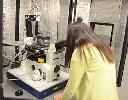
Model: Asylum Research MFP-3D Bio on top of a Nikon TE-2000
Capabilities: Height, conductive, magnetic, and fluorescent data
Operations Manual: Version 04.08 (18 MB)
Website: Asylum Research
X-ray Diffraction
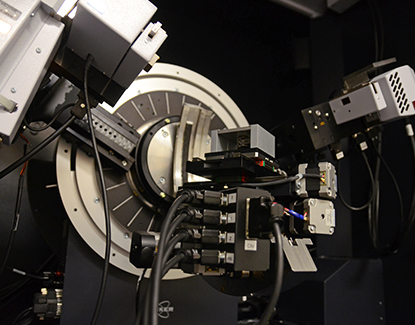
Model: Brucker D8 Discover
Scanning Electron Microscope with X-ray Detector EDS system
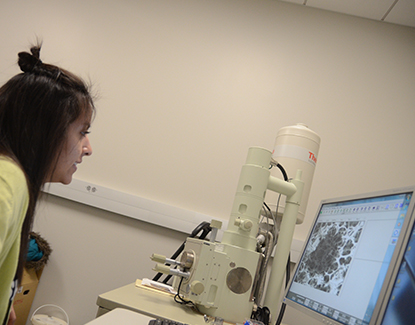
The scanning electron microscope (SEM) scans your sample with a beam of electrons to produce detailed information about the sample's topography and composition.
Model: JEOL 6060LV with Thermo Fisher Scientific NORAN System Six EDS
Capabilities: Imaging and X-ray data
Website: JEOL USA
Sputter Coater with Carbon Accessory for Thermal Deposition
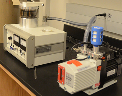
The sputter coater uses a sputter deposition process to coat your specimen with a thin layer of gold, silver, or a gold-palladium alloy, allowing it to be viewed in the scanning electron microscope (SEM).
Model: Denton Vacuum Desk IV
Capabilities: Gold, silver, and gold-palladium targets with thickness monitoring
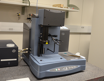
Differential Scanning Calorimeter (DSC)
Differential Scanning Calorimetry (DSC) is a thermoanalytical technique used to measure the difference in the amount of heat required to increase the temperature of a sample and a reference, as a function of temperature.
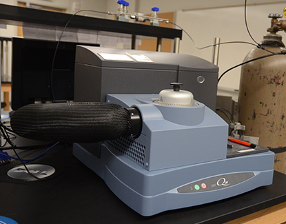
Thermogravimetric Analyzer (TGA)
Thermogravimetric Analysis (TGA) is commonly used to determine specific characteristics of materials that exhibit mass loss or gain due to decomposition, oxidation, or the loss of volatiles such as moisture.